What is the FluoroSpot assay?
The dual-color FluoroSpot assay is a modified T cell ELISPOT assay that enables the simultaneous detection of t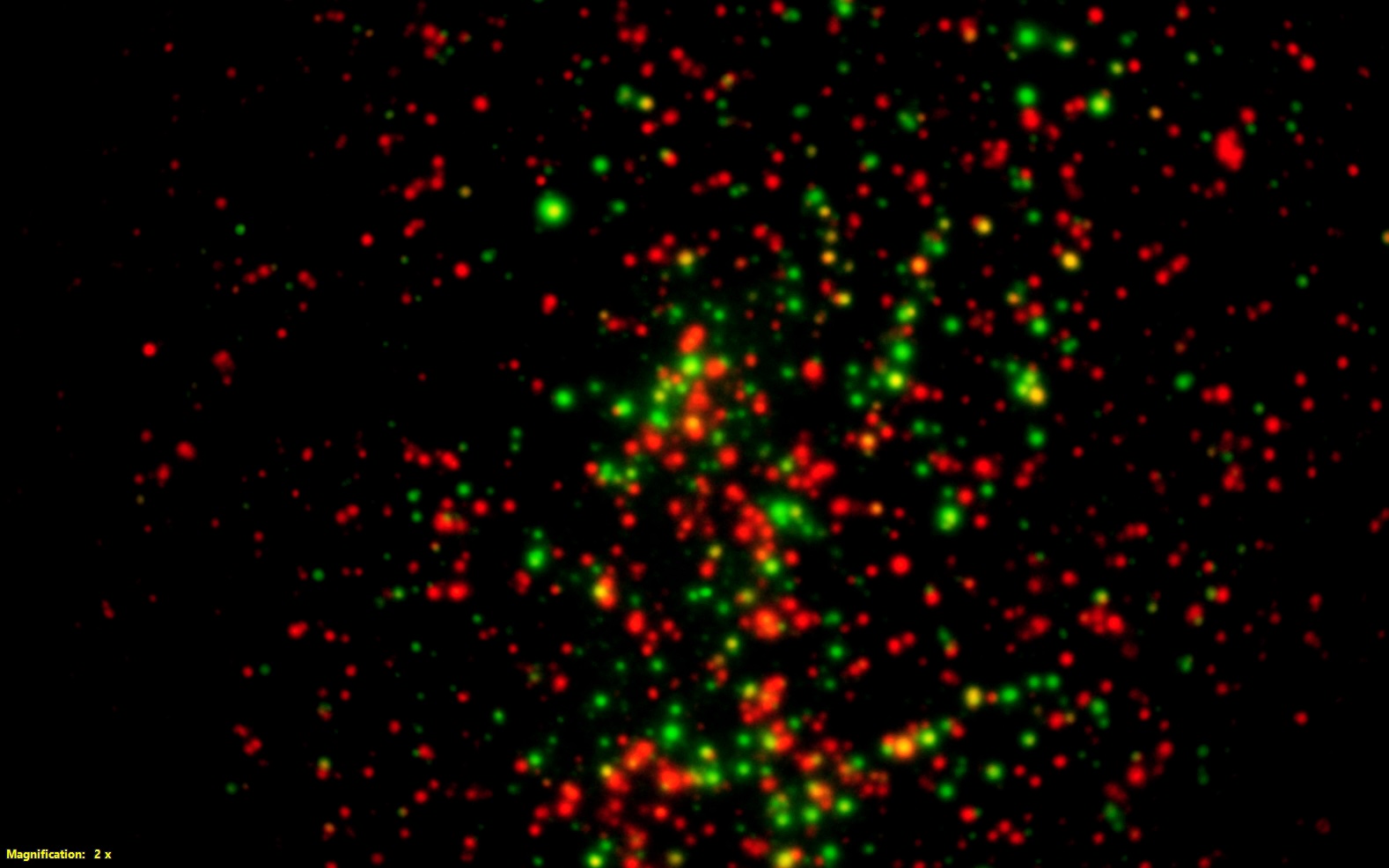 wo cytokines secreted by a single cell using fluorophore-labeled antibodies.
FluoroSpot is ideally suited for analyzing T cell subsets with distinct cytokine profiles, providing deeper insight into cellular immune responses.
The FluoroSpot assay is a highly sensitive method for detecting antigen-specific T cells and simultaneously measuring two cytokines in samples from immunized and non-immunized humans and animals.
Because the FluoroSpot assay detects two cytokines simultaneously, fewer PBMCs are required, making it particularly suitable for clinical studies and other applications where sample availability is limited.
Brief description of the FluoroSpot assay
The wells of a FluoroSpot plate are coated with two cytokine-specific capture antibodies. Cells are added and incubated, allowing secreted cytokines to bind to the corresponding capture antibodies. After incubation, the cells are removed by washing. Next, cytokine-specific detection antibodies and fluorescent conjugates are added.
Cytokine-producing cells are visualized as fluorescent spots. Green and red spots indicate cells producing a single cytokine, while yellow spots indicate cells producing both cytokines simultaneously.
Detection and visualization
Our FluoroSpot kits use a fluorescent conjugate system consisting of Alexa Fluor™ 488-labeled anti-FITC antibodies and R-phycoerythrin (R-PE)-labeled streptavidin. A spot enhancer is included to increase fluorescence intensity and improve spot detection.
A fluorescence microscope or an automated ELISPOT/FluoroSpot reader equipped with fluorescence detection capabilities is required to visualize and analyze the spots.
Overview of commonly used cell stimuli for ELISPOT and FluoroSpot assays
The stimuli described in this link for T cells, monocytes, and dendritic cells can be used in T cell ELISPOT and FluoroSpot assays as positive controls to assess cell functionality and verify proper assay performance.
The stimuli described for B cells can be used in B cell ELISPOT assays to activate memory B cells and promote their differentiation into antibody-secreting cells.
The concentrations listed should be considered as general guidelines.
It is recommended to evaluate a range of concentrations to determine the optimal stimulation conditions for your ELISPOT assay.
Up to 3×10⁵ cells can be added per well in a 96-well ELISPOT plate. However, the number of cells should not be so high that spot counting becomes difficult. For reliable analysis, a spot count of approximately 50 to 100 spots per well is generally recommended.
How should I dry the plate after color development?
PVDF membrane-bottomed plates should be dried without the lid and placed upside down at room temperature (20 to 26 °C), protected from light. It is important that the membranes are completely dry before the plates are evaluated.
Once dried, the plates should be stored in a dry place at room temperature, protected from light, to preserve fluorescence and prevent spot bleaching.
Storage of FluoroSpot plates
Fluorophores can lose their fluorescence when exposed to light, a process known as photobleaching.
To preserve fluorescence signal intensity and assay performance, FluoroSpot plates should be protected from light and moisture during storage at room temperature (20 to 26 °C).
How soon will FluoroSpot assay spots fade?
FluoroSpot assay spots are susceptible to photobleaching. When a fluorophore is exposed to light of the appropriate wavelength, it is excited from its ground state to an excited state. During this process, interactions with other molecules can lead to permanent chemical modifications that reduce fluorescence intensity. As a result, all fluorophores will gradually lose their fluorescence over time.
However, when FluoroSpot plates are stored in a dry place and protected from light at room temperature (20 to 26 °C), the spots remain relatively stable for several weeks to months.
For optimal results, we recommend evaluating FluoroSpot plates within one week after completion of the assay.
Please note that repeated exposure to light during analysis, for example while adjusting microscope settings or configuring an automated reader, can also accelerate photobleaching.
Which type of device is required to evaluate an ELISPOT or FluoroSpot plate?
For spot counting on PVDF membrane-bottomed ELISPOT plates, a reflected-light microscope or an automated ELISPOT reader is required. For FluoroSpot plates, a fluorescence microscope or an automated ELISPOT/FluoroSpot reader equipped with multicolor optical filters is required.
The reader should support detection of:
- FITC/Alexa Fluor™ 488 (Excitation/Emission: 495/519 nm) for visualization of green spots.
- R-Phycoerythrin (R-PE) (Excitation/Emission: 546/578 nm) for visualization of red spots.
Dual cytokine-producing cells appear as yellow spots when the green and red fluorescence signals overlap.
Why do I see high background or artificial spots in my FluoroSpot wells?
High background, diffuse fluorescence, or artificial spots in FluoroSpot wells can result from several factors, including inadequate washing, nonspecific antibody binding, contaminated reagents or cell cultures, carryover of secreted cytokines from a preincubation step, or incomplete membrane drying before analysis. Excessive cell numbers, overly long incubation times, use of non-recommended PVDF plates, incorrect reader settings, and autofluorescent dust particles can also contribute to background signals or interfere with spot detection.
To minimize background, follow the assay protocol carefully, use clean reagents and equipment, wash cells thoroughly after preincubation, ensure complete membrane drying, optimize cell density and incubation times, use the supplied FluoroSpot plates, and verify reader settings before analysis.
My question is not listed here. Where can I find more answers?
If you cannot find the answer to your FluoroSpot question in this section, please also refer to our ELISPOT FAQs. Since FluoroSpot is based on the ELISPOT assay principle, many topics such as sample preparation, assay optimization, plate handling, and data analysis are covered in the ELISPOT FAQ section and may provide the information you need.
If your question remains unanswered, please contact our customer service team at cs@ucytech.com.