FluoroSpot assay
The dual-color FluoroSpot assay, a modified T cell ELISPOT assay, enables simultaneous detection of two cytokines released from a single cell using fluorophore-labeled antibodies.
The FluoroSpot assay is ideal for the analyzing T cell subsets with with distinct cytokine profiles.
U-CyTech biosciences offers a broad selection of human and monkey cytokine FluoroSpot kits, including dual detection kits for IFN-γ and a secondary cytokine (e.g. IL-2, IL-10, IL-13, IL-17A). Our monkey FluoroSpot kits cross-react with various macaque and baboon species. Each kit includes all necessary reagents and PVDF membrane-bottomed (PVDF) plates optimized for the FluoroSpot assays.
Application
- The FluoroSpot assay is a highly sensitive assay for detecting antigen-specific T cells and simultaneously measuring two cytokines in immunized and non-immunized humans and animals.
- Because the FluoroSpot assay detects two cytokines simultaneously, it requires fewer PBMCs, making it ideal for clinical trials with limited sample availibility.
- The FluoroSpot assay can assess T cell responses in individuals with type 1 diabetes at the single-cell level
 (Arif S. et al. 2022).
Brief description of the FluoroSpot assay
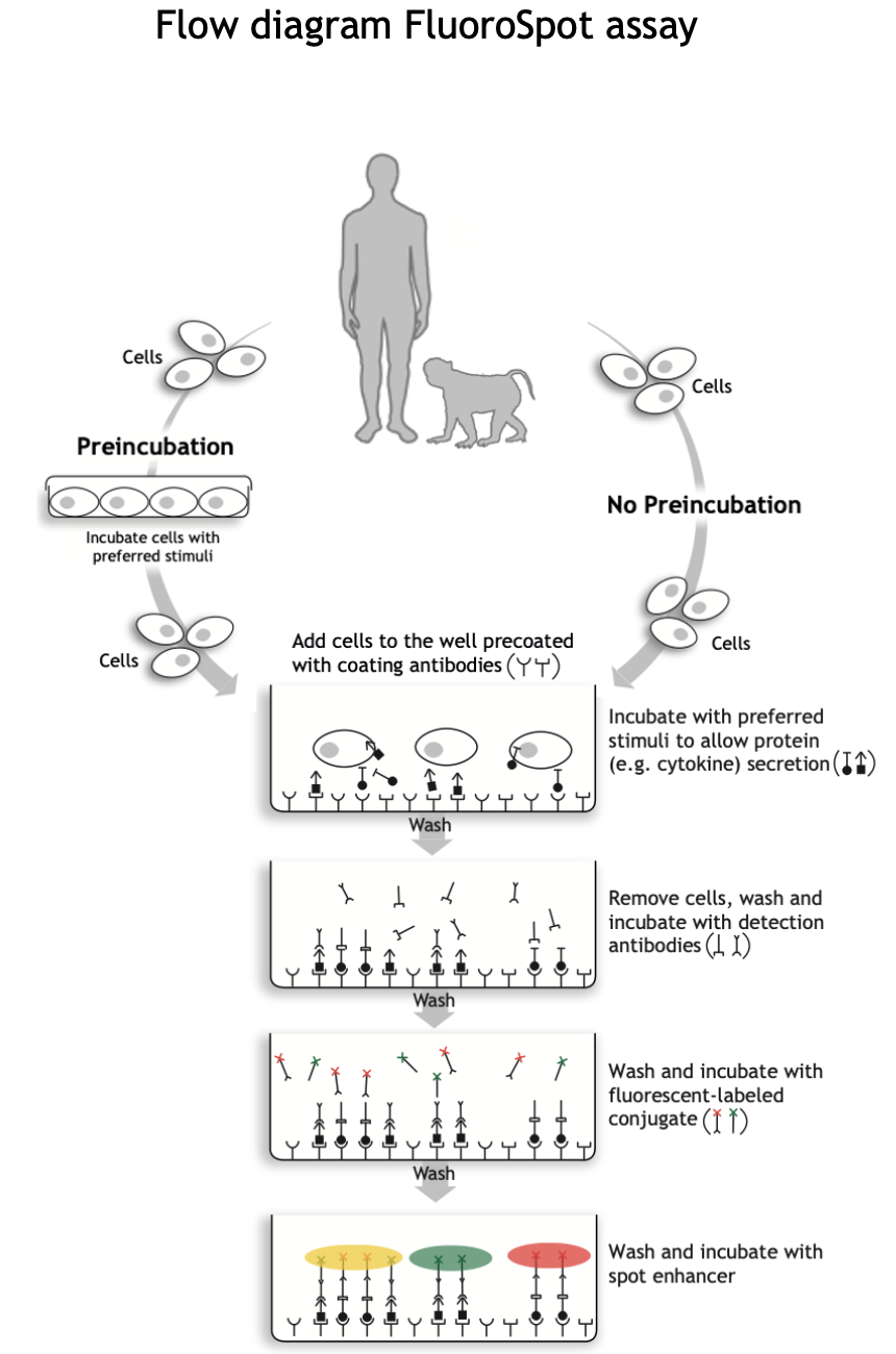
Cells are incubated in the wells of the FluoroSpot plate coated with two cytokine-specific cytokines. After binding of the released cytokines, the cells are washed away. Detection antibodies followed by two fluorescent labeled conjugates are added.
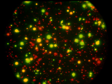
Green and red fluorescent spots show single cytokine producers, while yellow spots produce both cytokines.
The different steps of the FluoroSpot procedure are illustrated in the FluoroSpot assay Flow diagram.
U-CyTech FluoroSpot products
Follow these links to go to directly our: